
About
Putting 3D in-vitro research within reach of any lab
ALIOS-PES delivers an integrated hardware, software and biomaterial portfolio for suspended 3D bioprinting — so a laboratory can produce reproducible 3D tissue models without a dedicated bioprinting specialist.
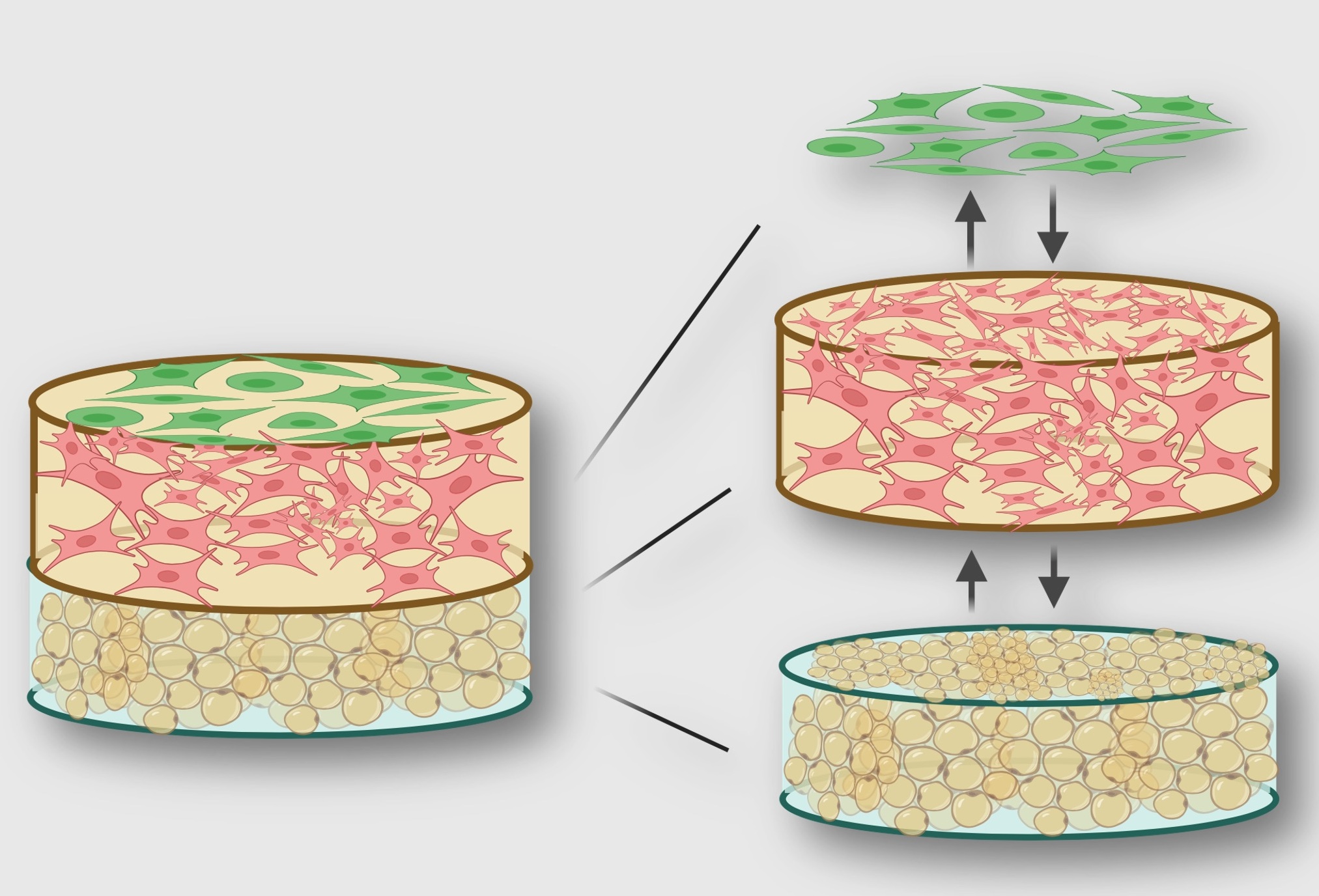
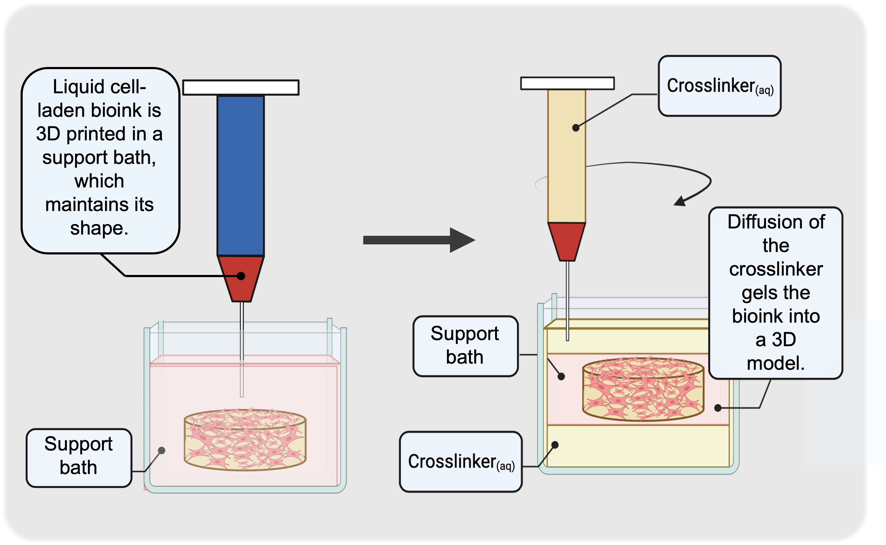
The science: suspended layer additive manufacture
Suspended 3D bioprinting uses a quasi-liquid support environment to hold cell-laden biomaterials in place as they transition from liquid into hydrogel bioscaffolds — however long that gelation takes.
Decoupling shape retention from cure time enables the sequential printing of distinct layers and features — each offering full control over biomaterial composition, the embedded cell type and cell density. The technology further can accommodate a broader range of bioinks, compared to traditional extrusion approaches. That is the basis of every product in our portfolio.
Validated applications
All presented applications have been produced with a development version of the Polysaccharide–Collagen I reagent kit.
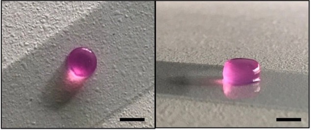
The technical replicate
Available for 3D cell culture on its own, or as a layer in a multi-layer co-culture in-vitro 3D tissue model. Compatible with long-timeline studies of 21 days and over.
Scale bar: 5 mm.
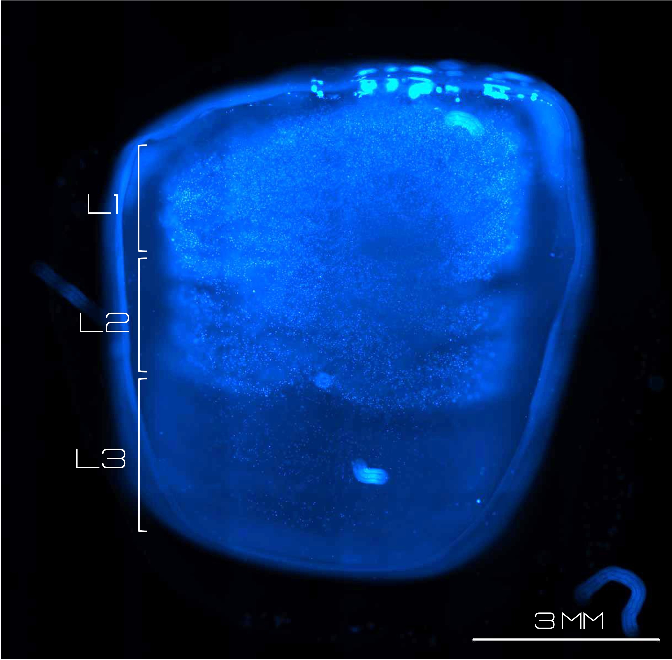
Multi-layer integration
Integration of distinct layers in a single technical replicate for complex tissue in-vitro modelling. Each layer is seeded at 0.5×10⁶ cells/ml (bottom layers), 1.0×10⁶ cells/ml (middle layer) and 1.5×10⁶ cells/ml (top layer).
Images show cross-sections stained with Hoechst 33342 nuclear stain. Scale bar: 3 mm.
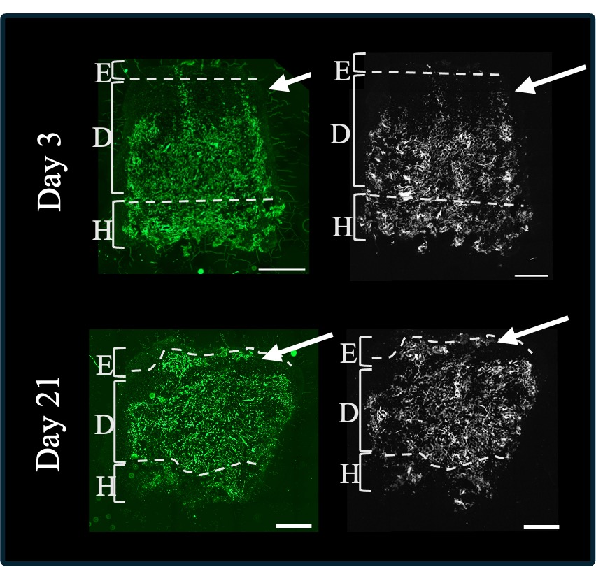
Label-free ECM characterisation
Label-free characterisation for insight with minimal processing. Fluorescent (green) and second harmonic generation (B&W) imaging showcasing ECM deposition at the dermal–epidermal junction (white arrow) of full-thickness in-vitro 3D skin models after 21 days of culture, compared to 3 days. Models composed of human epidermal keratinocytes, human dermal fibroblasts and adipose-derived stem cells.
Legend: E, D and H — epidermis, dermis and hypodermis, respectively. Scale bar: 1 mm.
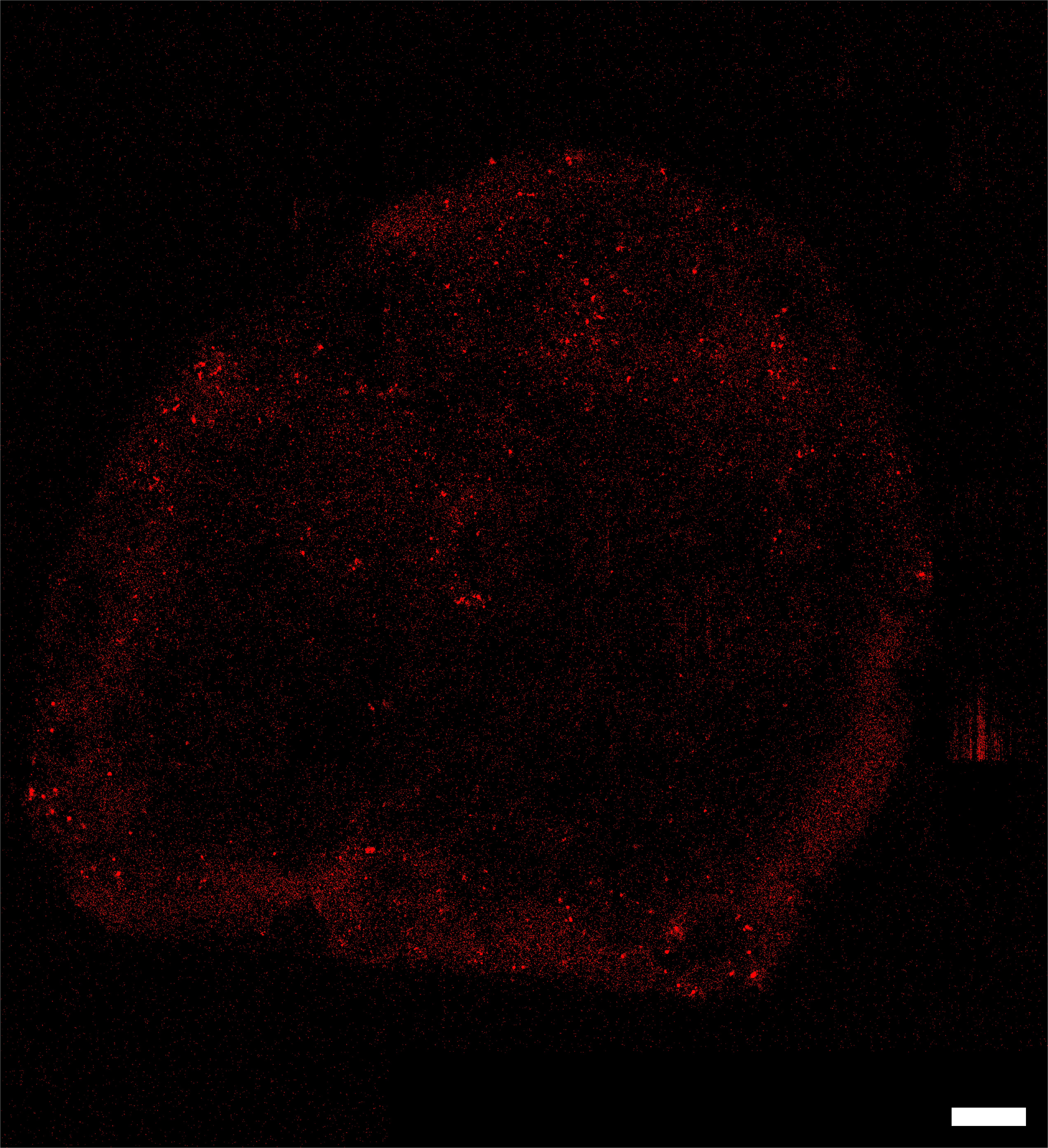
Immunohistochemistry in bulk sections
Avoiding the harshness of standard microtomy prep, local protein expression can be resolved within a bulk 800 µm section of a bioprinted construct, only fixed with 4% Paraformaldehyde. Here, α-smooth muscle actin (SMA) is visualised in a full-thickness in-vitro 3D model of skin, comprised of a hypodermis, a dermis and an epidermis after 21 days' culture.
Scale bar: 1 mm.
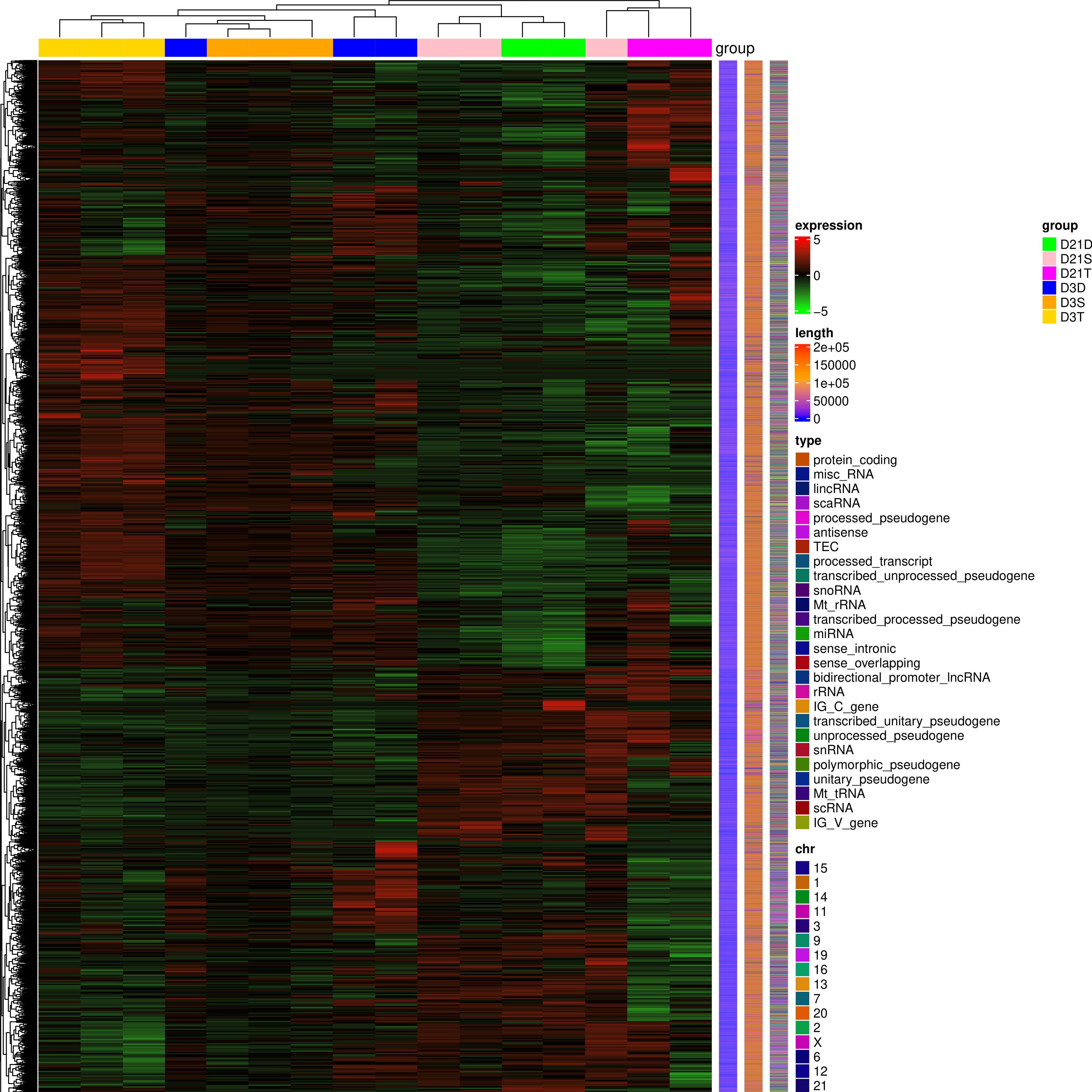
Full-transcriptome RNAseq
Full-transcriptome RNAseq for comprehensive analysis of in-vitro phenotypes. The image presents a section of the heat map from a study on the development of in-vitro skin in full-thickness 3D models, comparing bulk model transcription profiles after 3 days and 21 days of culture.
Trusted by
